The AOSR uses cookies on its site to make your browsing experience better. By clicking OK, you agree to our terms of use. To find out more, visit our cookie policy.

| Chief complaint | Ankle sprain with pain. |
|---|---|
| Age | 62yrs |
| Sex | Male |
| Modality | CT,Plain film/Xrays |
| System | Musculoskeletal |

Kwong Wah Hospital
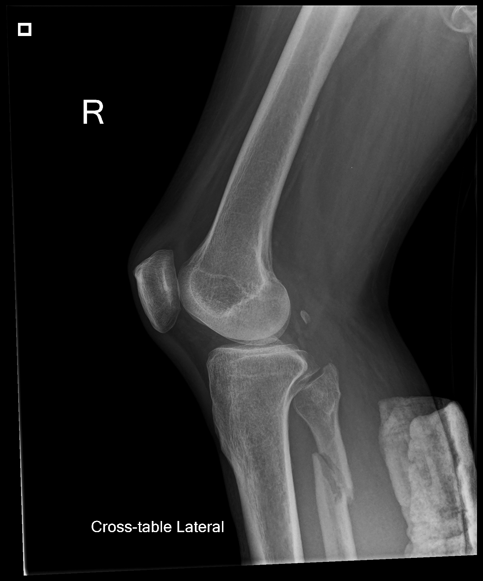
Maisonneuve Fracture
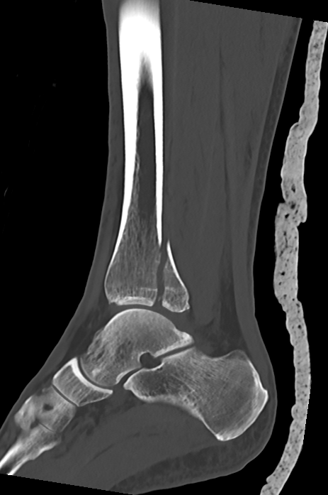
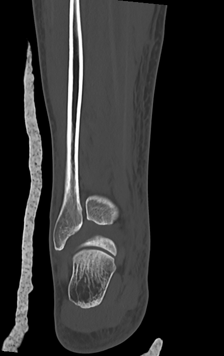
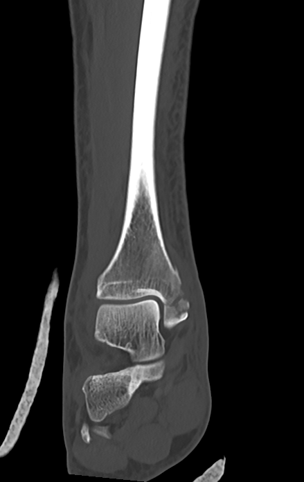
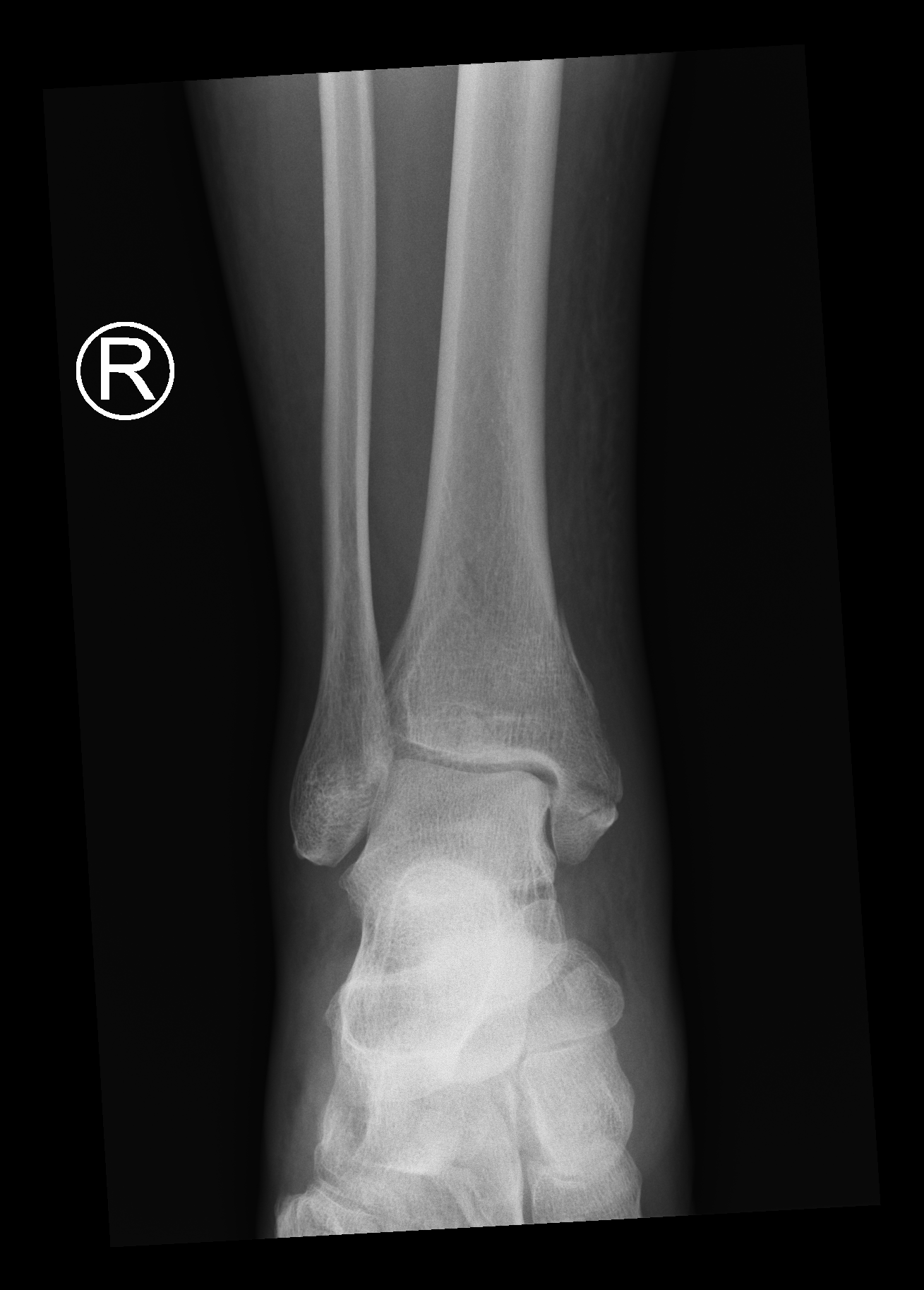
XR and CT right ankle show medial malleolar multi-fragmentary fracture with intra-articular extension. Posterior malleolar fragment involves >25% of anteroposterior tibial plafond diameter. Distal fibula in scan range shows no fracture. While there is no significant widening of distal tibiofibular syndesmosis or mortise joint, the extent distal tibial fracture prompts for further assessment of the entire fibula. Subsequent XR show proximal fibula fracture.
Overall findings are compatible with Maisonneuve fracture (Lange-Hansen pronation-external rotation PER stage IV, Weber C, pilon variant). Maisonneuve fracture describes a combination of proximal supra-syndesmotic fibula fracture with unstable ankle injury, which can present as injury to deltoid or distal tibiofibular syndesmosis, or with medial malleolar fracture. It occurs through pronation-external rotation of ankle during injury. Maisonneuve fracture usually require surgical management.
This case highlights the importance of imaging the entire fibula when medial malleolar fracture or evidence of ankle instability (including widened mortise or distal tibiofibular syndesmosis) is present without lateral malleolar fracture.
Reference
1. Bartoníček J, Rammelt S, Kašper Š, Malík J, Tuček M. Pathoanatomy of Maisonneuve fracture based on radiologic and CT examination. Archives of Orthopaedic and Trauma Surgery. 2019 Apr 1;139:497-506. DOI: https://doi.org/10.1007/s00402-018-3099-2
2. Hanson JA, Fotoohi M, Wilson AJ. Maisonneuve fracture of the fibula: implications for imaging ankle injury. AJR. American journal of roentgenology. 1999 Sep;173(3):702. DOI: https://doi.org/10.2214/ajr.173.3.1047090.