The AOSR uses cookies on its site to make your browsing experience better. By clicking OK, you agree to our terms of use. To find out more, visit our cookie policy.

| Chief complaint | Presented with occipital headache and double vision for past five weeks. |
|---|---|
| Age | 64 yrs |
| Sex | Male |
| Modality | CT/MRI |
| System | Head and Neck |

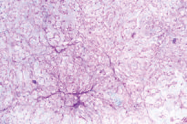
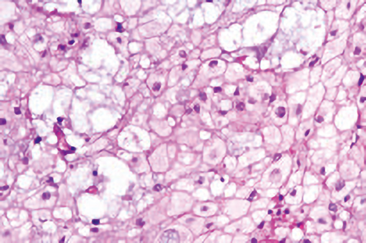
The histo-pathological findings confirmed the diagnosis of sellar chordoma.
Sellar chordomas are extremely rare and are often misdiagnosed as pituitary adenoma. The lesion in above case is markedly hyperintense on T2WI (attributed to the high fluid content of vacuolated cellular components) and showed heterogeneous enhancement with osseous erosion, which favored possibility of malignant chondroid lesion. In contrast, pituitary macroadenoma show intermediate signals on T2WI. The other T2 hyperintense lesions include dermoid and epidermoid cysts, however they are non-enhancing. No heterogeneity was noted on T1WI to suggest proteinaceous component and hence craniopharyngioma and Rathke’s cleft cyst were also excluded.