The AOSR uses cookies on its site to make your browsing experience better. By clicking OK, you agree to our terms of use. To find out more, visit our cookie policy.

| Chief complaint | A six-day old baby was delivered through meconium-stained liquor. The baby did not cry after birth and developed respiratory distress soon after and was started on nasal cannula flow. With progressive tachypnea, increasing FiO2 requirement and hypercarbia, the baby was electively intubated and connected to mechanical ventilator. Since echo showed features of a vascular ring, CT angiogram of chest was done. |
|---|---|
| Age | Six days |
| Sex | N/A |
| Modality | CT |
| System | Chest |

Amrita institute of medical sciences
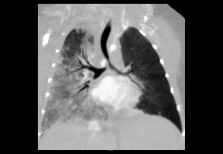
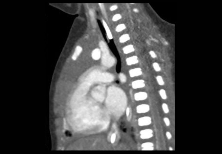
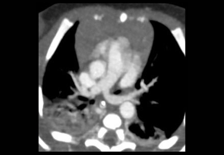
Aberrant origin of left pulmonary artery is
noted from the right pulmonary artery. It is thereafter seen to course between
the esophagus (represented by nasogastric tube) and the distal trachea and
carina, causing airway compression.
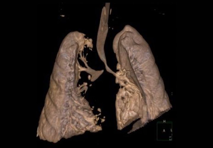
Aberrant left pulmonary artery, also known
as pulmonary sling, represents an anatomical variant characterized by the left
pulmonary artery arising from the right pulmonary artery and passing above the
right main bronchus and in between the trachea and esophagus to reach the left
lung. It may lead to compression and focal stenosis of the trachea. Compared to
other vascular rings from aortic anomalies which run behind the esophagus, pulmonary
vascular sling anomaly course between trachea and esophagus.
The formation of a pulmonary sling (anomalous left pulmonary artery) is the result of a failure of the proximal left 6th arch to properly involute. An anastomotic vessel, connecting the primitive pulmonary circulations, becomes the anomalous left pulmonary artery, arising from the right pulmonary artery. This vessel then travels above the main pulmonary bronchus to reach the left lung hilum by passing between the trachea and esophagus, often leading to a compression of these structures. They were found to make up approximately 4% of congenital vascular anomalies. Common presenting complaints are wheezing, stridor, vomiting and feeding difficulties
References:
1. Newman B, Cho Y. Left pulmonary artery
sling—anatomy and imaging. Semin Ultrasound CT MR. 2010; 31:158-170
2. Grover FL, Norton JB, Jr, Webb GE,
Trinkle JK. Pulmonary sling. Case report and collective review. J Thorac
Cardiovasc Surg. 1975; 69:295–300