The AOSR uses cookies on its site to make your browsing experience better. By clicking OK, you agree to our terms of use. To find out more, visit our cookie policy.

Quiz 5

Test your knowledge, take our self-directed learning Quiz.

Case information
Chief complaint A 1.5-year-old boy was referred to a paediatric surgeon with a 3-month history of left submandibular mass. The kid is playful with no constitutional symptoms. On physical examination, there was a ~3cm soft, non-tender and mobile mass at left submandibular region. The surgeon requested a contrast-enhanced MR scan for further evaluation.
Age 1.5 year
Sex Male
Modality MRI
System Head and Neck
Case contributor
Dr. Milly Chiu

Hong Kong Children's Hospital

Dr.Kevin Fung

Hong Kong Children's Hospital

The answer is
Imaging findings

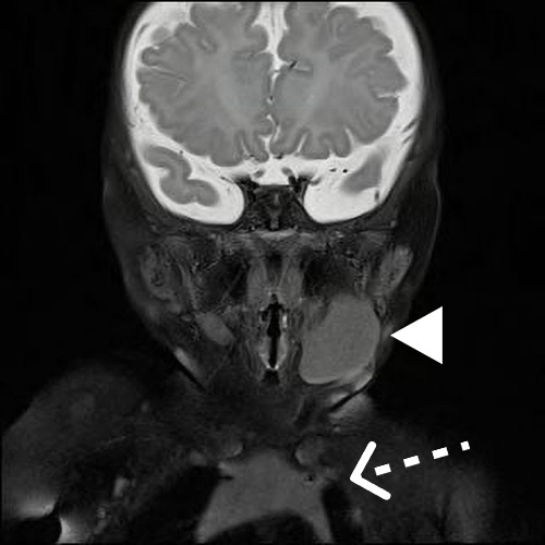
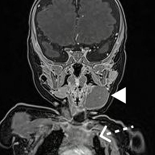
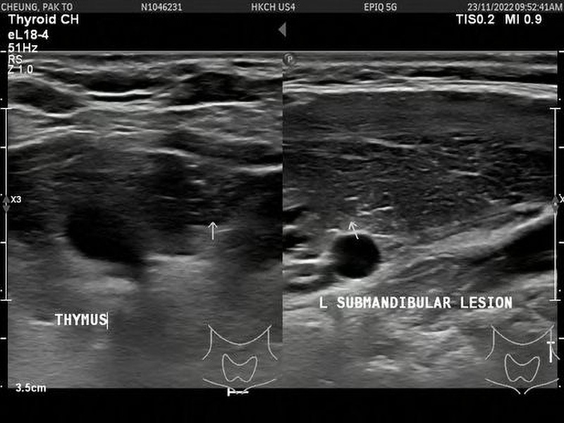
The MR scan showed a well circumscribed homogenous mass posterior to left submandibular gland. One key diagnostic clue was that the signal intensity and enhancement pattern of this mass (arrowhead) was IDENTICAL to that of the thymus (dashed white arrow).

Discussion

Embryologically, the thymus develops from the 3rd branchial pouches bilaterally. Towards the end of the 6th week, the thymic lobes on each side traverse caudomedially into the superior mediastinum before fusing in the midline. Ectopic tissue may occur anywhere along this path secondary to abnormal descent, implantation or persistence of remnant tissue, resulting in a thymus positioned in the neck above the brachiocephalic veins. It is important to recognise this entity and NOT to mistake it as malignant neck tumours. Ultrasound is the best investigation to demonstrate the characteristic sonographic pattern of the thymus. Biopsy in these children are unnecessary and should be avoided.

The classic description is a "starrysky appearance", which describes multiple speckled echogenic foci (white arrow)on a background of homogenous hypoechoic parenchyma.

The echogenic foci represent the fine septae within the normal thymus gland.


Reference:

Chu, W.C.W. and Metreweli, C. (2002), Ectopic thymic tissue in the paediatric age group. Acta Radiologica, 43: 144-146.