The AOSR uses cookies on its site to make your browsing experience better. By clicking OK, you agree to our terms of use. To find out more, visit our cookie policy.

| Chief complaint | A 73-year-old man was sent for evaluation of chronic diffuse abdominal pain accompanied by anorexia and weight loss. Look at the USG followed by CT to determine the origin and probable diagnosis. |
|---|---|
| Age | 73 |
| Sex | Male |
| Modality | CT |
| System | Abdomen,Chest,Vascular |

India
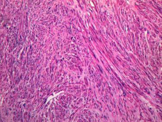
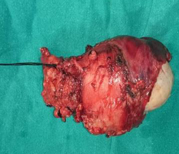
There is a large lobulated heterogeneously enhancing intraluminal Inferior Vena Cava mass with necrotic changes in the suprarenal inferior vena cava distending and obliterating the lumen. There is no invasion into hepatic parenchyma. Left renal vein is distended with hypodense non enhancing thrombus. Right renal vein shows optimal luminal opacification. Color Doppler examination reveals no flow into left renal vein. Juxtahepatic Inferior vena cava shows optimal luminal caliber and contrast enhancement.
Bland Thrombus in Inferior Vena Cava: There is enhancing intraluminal mass with gross expansion of the inferior vena cava with no flow channels in or around the mass, which are features of tumoral thrombosis.
Paracaval/Caval Lipoma: The mass shows heterogeneous enhancement with no obvious fat density attenuation values.
Adrenal Mass: Right adrenal gland is seen separate from the lesion with normal size, shape and enhancement characteristics.
Renal Cell Carcinoma: Right kidney is visualised distinct from the lesion with normal parenchyma.